Posts

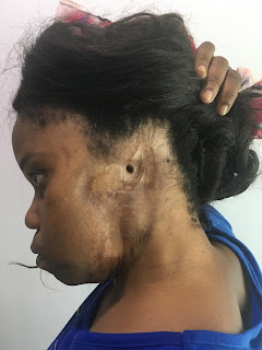
Loss of Ear - First stage Microvascular Radial forearm flap
Image
This Lady lost her left ear to a road traffic accident in Zambia. Referred to us for Reconstruction of ear. As a part of reconstruction the scarred area around the external auditory meatus is replaced with microvascular radial forearm free flap with a small opening the edges of which were sutured with the external auditory meatus to maintain external auditory meatal opening. Ear reconstruction with Firmin technique will b planned once the flap is settled. 
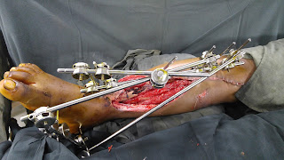
Crush Leg - Anterolateral thigh flap

Image
Crush injury of the leg with multiple communited fractures of Both bones of leg (compound grade3b). After Debridement and stabilization of wound, Anterolateral thigh flap was harvested from the thigh and the wound on the leg was covered. An end to side anostomosis was done here with Posterior tibial artery because anterior tibial artery gave a surpise during surgery in form of contusion. Flap well healed.

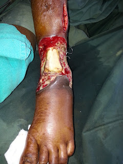
Latissimus doors free flap for exposed ankle

Image
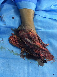
Post traumatic runover leading to exposed ankle joint Covered with free latissimus dorsi flap. Latissimus dorsi muscle was selected due to risk of infection. Patient was shifted to our hospital with sepsis due to this wound which we improved.

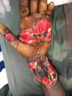
Electrical Burns Entry wound - Anterolateral thigh free flap

Image
Electrical burns leading to exposed vital structures of wrist. Anterolateral microvascular flap was used to cover the wrist. 

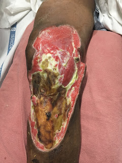
Necrotisisng fascitis defect - Free Latissimus dorsi flap

Image
Necrotising fascitis of knee leading to loss of skin and subcutaneous tissue with exposed knee and its ligaments. After debridement and wound stabilization stable wound cover was done with Microvascular Latissimus dorsi muscle flap.

Crush foot with Anterolateral thigh flap

Image
Crush foot  Microvascular Anterolateral thigh flap Reconstruction with Skeletal fixation

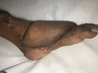
Foot and Ankle reconstruction - Free Latissimus dorsi Musculocutaneous flap

Image
Crush injury of the foot due to runover by van Initial Skeletal stabilisation and debridement done. Because of preserved vascularity decision to reconstruct the foot was taken. Microvascular Latissimus dorsi musculocutaneous flap was done.