Posts

Showing posts from September, 2019

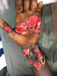
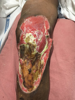
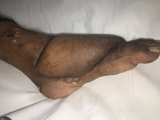
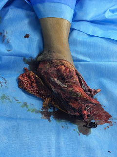
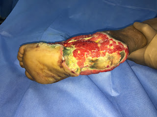
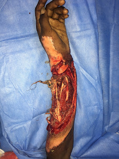
Latissimus doors free flap for exposed ankle

Image
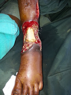
Post traumatic runover leading to exposed ankle joint Covered with free latissimus dorsi flap. Latissimus dorsi muscle was selected due to risk of infection. Patient was shifted to our hospital with sepsis due to this wound which we improved.

Electrical Burns Entry wound - Anterolateral thigh free flap

Image
Electrical burns leading to exposed vital structures of wrist. Anterolateral microvascular flap was used to cover the wrist. 

Necrotisisng fascitis defect - Free Latissimus dorsi flap

Image
Necrotising fascitis of knee leading to loss of skin and subcutaneous tissue with exposed knee and its ligaments. After debridement and wound stabilization stable wound cover was done with Microvascular Latissimus dorsi muscle flap.

Crush foot with Anterolateral thigh flap

Image
Crush foot  Microvascular Anterolateral thigh flap Reconstruction with Skeletal fixation

Foot and Ankle reconstruction - Free Latissimus dorsi Musculocutaneous flap

Image
Crush injury of the foot due to runover by van Initial Skeletal stabilisation and debridement done. Because of preserved vascularity decision to reconstruct the foot was taken. Microvascular Latissimus dorsi musculocutaneous flap was done.

Crush injury Heel and foot - Free Latissimus dorsi flap with skin graft

Image
Crush injury of left foot with loss of heel skin and fat pad with loss of skin over dorm.  Free Latissimus dorsi flap was planned and Colour doppler of the left lower limb was done to screen for vascularity and was normal. CT- Angiography couldn't be done to raised creatinine levels. Intraoperatively Anterior tibial vessels were found to be hypoplastic. Posterior tibial vessels were hardened and atherosclerotic. An End to side anastomosis was done with posterior tibial artery.  Patient was kept on blood thinners for 3 months to prevent thrombosis. A well healed flap with normal function of the limb achieved.

Anteromedial thigh flap

Image
Crush injury left forearm with vascular injury and Radius fracture (Grade IIIb). Initial debridement with vascular repair and External fixator application done. Anteromedial thigh microvascular flap done to cover all the vital structures.  Initially Anterolateral thigh flap was planned and after confirming and marking the perforator preoperatively by doppler surgery was posted. Intraoperativey Perforator was too thin and too distal. The doppler signal preoperatively must have been a muscle perforator that is not communicating with the draining vessel Intraoperatively Perforator for the Anteromedial thigh flap was found to be robust and plan was improvised to Anteromedial thigh flap. A well settled flap post operatively.

Mandibular Reconstruction Carcinoma Buccal Mucosa - Fibula Composite Free flap

Image
Carcinoma Left GB Sulcus with involvement of mandible and retromolar trigone. Hemi mandibulectomy done for the same Mandible was primarily reconstructed with Microvascular fibula bone flap. Post operative X-rays suggest of well integrated bone.